

0 7567 3000
0 7567 3000
Walailak Frontier 11, January 2019
Imaging can be thought as a way to visually construct an object. The technique is in general based on the use of electromagnetic waves ranging from gamma ray to radio wave. A variety of physical information can be extracted from the image either quantitatively or qualitatively based on their appearance, e.g., size and dimensions of objects of interest, and their visual characteristics. However, mechanical characteristics such as elastic modulus of or vibrational attenuation within the object are unavailable. To fill the missing gap, imaging with picosecond ultrasonics comes into play. It gives the insight to those mechanical properties. Previously, the technique was widely used to determine thickness of thin film and explore thermal properties of condensed matter. Until recently, the technique has been applied to study mechanical characteristics of biological tissues.
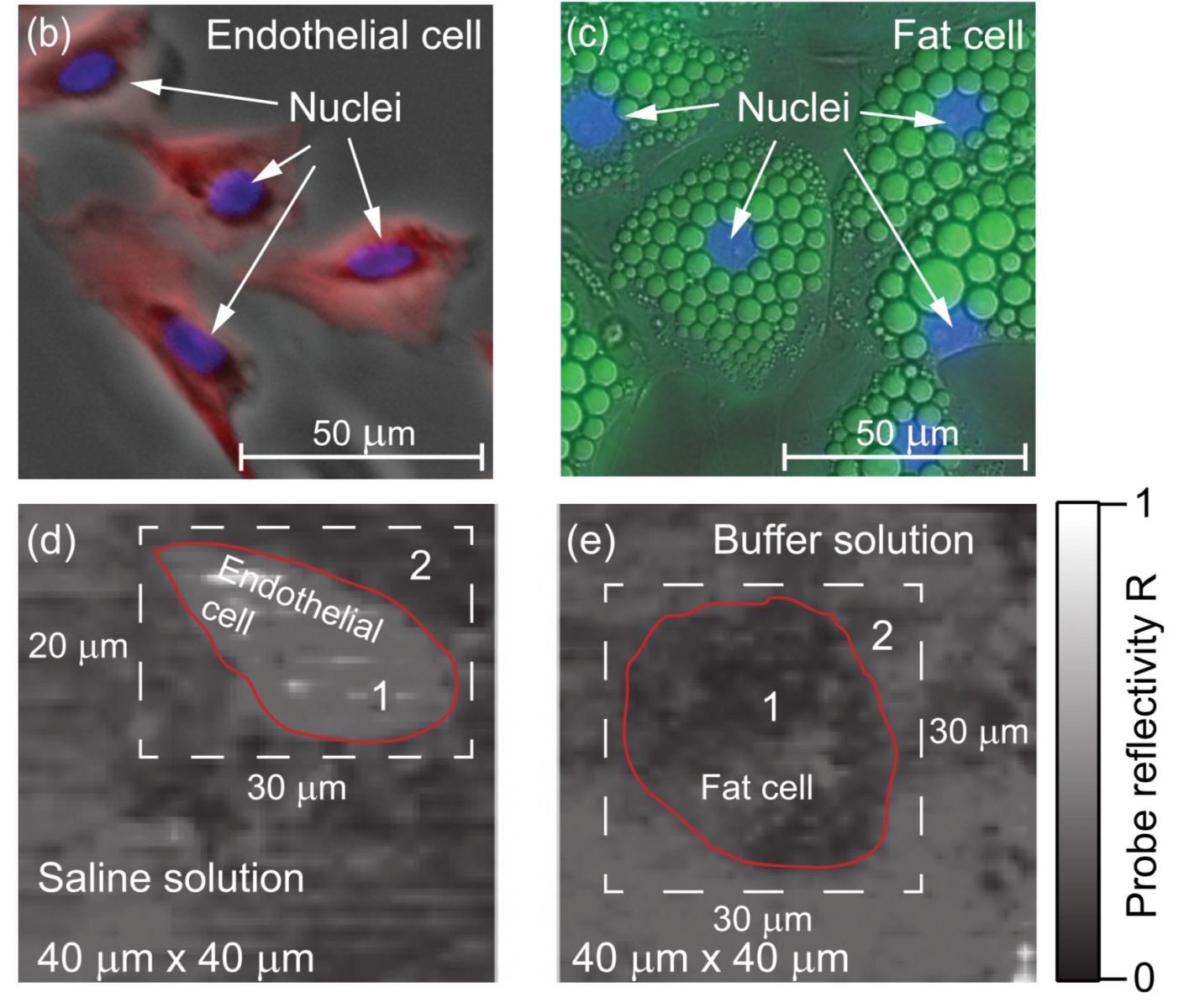
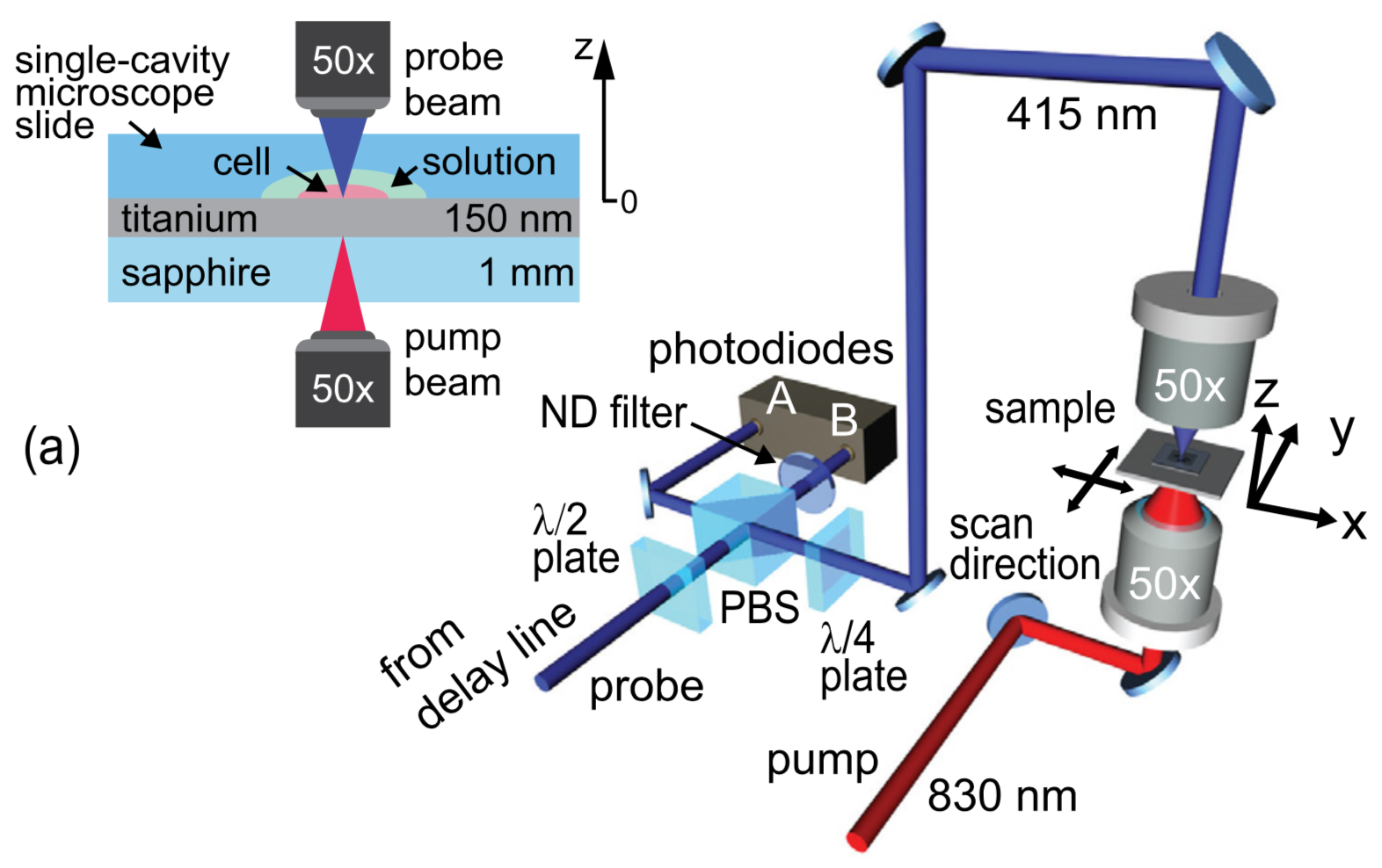
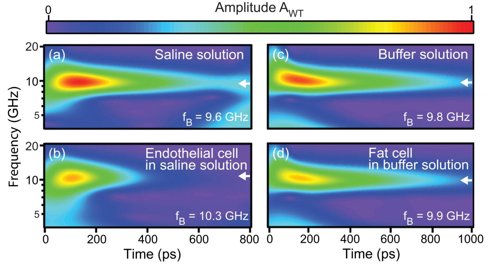
We use picosecond ultrasonics to image animal cells in vitro-a bovine aortic endothelial cell and a mouse adipose cell. They were fixated to Ti-coated sapphire. Tightly focused ultrashort laser pulses generate and detect GHz acoustic pulses, allowing three-dimensional imaging (x, y, and t) of the ultrasonic propagation in the cells with 1-mm lateral and 150 nm depth resolutions. Time-frequency representations of the continuous-wavelet-transform amplitude of the optical reflectivity variations inside and outside the cells show GHz Brillouin oscillations, allowing the average sound velocities of the cells and their ultrasonic attenuation to be obtained as well as the average bulk moduli. This was done to prove the possibility of using picosecond ultrasonics for imaging biological cells in order to deduce physical information of the cell as a whole in one experiment.
The work was done by Assoc.Prof.Dr.Sorasak Danworaphong (a researcher of Walailak University) and his collaborators in Japan taking temporal series of rasterizing images from the pump-probe experiment. The images were processed using wavelet transformation that allowed us to identify when certain frequencies occurred. Knowing the propagating time of those frequencies helped locating where the acoustic pulse was inside the cell. The frequencies also implied the acoustic responses of cell components since each component in the cell has different acoustic impedance. From this information, we were able to extract bulk modulus, sound speed, temporal and spatial attenuation of acoustic waves within the tissues.
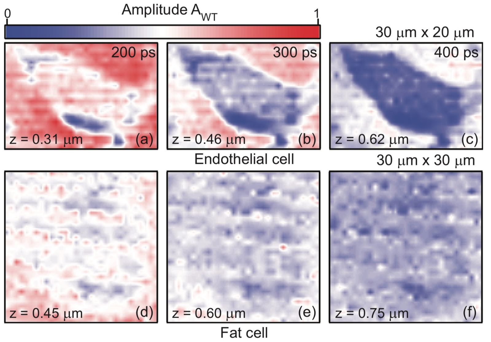
We obtained the average sound velocities, ultrasonic attenuation, and average bulk moduli of the cells. All values were in close agreement with those obtained from other methods. The result gave stacks of ultrasonic images representing cells in z-direction or cell thickness. This is similar to peeling the cell layer by layer, allowing us to explore each layer individually. The technique could be improved in resolution by using shorter wavelength laser that can be focused more tightly. The substrate could be also replaced with higher heat conductive material. It helps dissipating heat faster that is very useful when higher power laser is used to improve image contrast. The cell would not be damaged from the lingering heat on the substrate.
This study, we measured the level of Alu methylation in normal, pre-type 2 DM, and type 2 DM patients by ALU-Combined Bisulfite Restriction Analysis . We investigate Alu methylation levels of white blood cells of type 2 DM, pre-DM, and control . The DM group possess the lowest Alu methylation (P < 0.001, P < 0.0001 adjusted age ). In the DM group, Aluhypomethylation is directly correlated with high fasting blood sugar, HbA1C, and blood pressure. Therefore, genome-wide hypomethylation may be one of the underlining mechanisms causing genomic instability in type 2 DM. Moreover, Alu methylation levels may be a useful biomarker for monitoring cellular senescence in type 2 DM patients.
Sources:
Facebook: wufrontovation
Related Link: